Nanoscience Photography
Photographer Felice Frankel captures phenomena invisible to the naked eye.
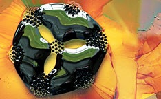
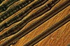
Ferrofluid
“Many scientists take pictures of nanoscience, but the pictures don’t really communicate what is going on,” says Felice Frankel. “I wanted people to see how fascinating that world is.”
No small task, even for her. Often called a “photoscience journalist,” Frankel, 64, has spent the past 19 years doing photography that’s not just beautiful but illustrates scientific concepts. She was an architecture shooter when, on a Loeb Fellowship to Harvard, she audited a class with chemist George C. Whitesides. He invited her to try to make an image for one of his articles, on hydrophobic surfaces. Her resulting “Chiclets” photo of water droplets ran on the cover of Science.
But her latest book with Whitesides, No Small Matter: Science on the Nanoscale ($35, Harvard University Press), was a new challenge. She had to capture subjects as tiny as DNA, ribosomes, and quantum dots— plus things that can’t be photographed literally, such as the principle of quantum mechanics. “The concept came first,” she says. “It was an entirely different thought process.”
For a chapter on music, Frankel mounted her Nikon D3x body on a microscope to capture how audio waves are imprinted on a Beatles record. She scanned small sea creatures and fuel cells at 600 dpi on a flatbed scanner. Close-up images of water droplets required a macro lens and creative props. Occasionally she’d rely on metaphor, using Adobe Photoshop to create images of the impossible.
“It’s giving readers a visual cue to make sense of these concepts,” Frankel says. “And it’s making pictures of research interesting to look at.”
**Interview With Felice Frankel by **Lori Fredrickson:****
You are in an unusual category of photographer…
Nobody is really doing what I’m doing, which is what some people call a “photoscience journalist”. There are people who have taken photographs of science, of course, but they’ve specialized in certain areas—microscopic work in biology, for example. My work is more generalized. I’m creating images of research, which doesn’t look like that research for the most part, because they are interesting images to look at.
Can you tell me how you first got started in photography?
_Before I started photography, I was trained in science, and did research in the science field. And then my husband bought me a Nikon—a good camera to start off with—and I took it up as a hobby. At one point I volunteered some of my time to take promotional shots for a local public television station. They’d just moved to a new building, and when the architect who had designed it saw me with my camera, he asked me if I knew how to take architectural pictures. _
And I lied. I said, “Sure, I know how to do that.” And I didn’t at all! But then I gave it a shot, and it turned out that I loved taking pictures of architecture, and that I had an eye for it. It’s really kind of creating an order—two dimensions out of a three-dimensional thing. So I started making pictures of the new space as well as an outdoor space for him, and he liked them both. My images were then published in an article in the magazine Garden Design, which launched my professional career. I started getting hired by the magazine, and then worked for a number of firms, architectural and landscape design firms.
And so how did this lead to science photography?
At one point I took photographs for a book on architecture, and as a result of this, I was given a Loeb fellowship at Harvard. While I was there, I had the freedom to audit any kind of class I wanted. And because science is my other big interest, I found myself auditing every conceivable science class that I could.
One of these was by George M. Whitesides, the scientist who did the writing in No Small Matter. We began talking after class, and he invited me to his lab to look at what he was doing. He was working on a project on hydrophobic and hydrophilic surfaces, or types of water-repellant surfaces, and was just about to submit an article on it to Science magazine. I looked at the pictures he had for it and, frankly, they were lousy. I said, let me give it a shot. And the photograph I took ended up getting on the cover of the magazine, and opened up a whole new set of doors for me.
What was the photograph?
_The Science magazine cover was a surface that had areas in it that were both hydrophobic and hydrophilic. When you wax your car, you’re actually making a hydrophobic surface. They were controlling a particular surface. What I did was ask them to make an interesting pattern on the surface and then I just flopped some water on it so that you can see the pattern more clearly. _
_They call it the Chiclet cover because it looks like a series of blue and green Chiclets—squares that are four millimeters apart, with colored water placed on them. They’re literally square drops of color. The subject turned out to be a very important new technique to use certain kinds of devices. _
_Then I took more pictures, got more covers, then went to MIT and carved out a place there for a few years. George Whitesides and I collaborated on a previous book also, entitled On The Surface of Things. The beauty of it is that I’m constantly learning. It’s not just making pictures. I’ve got to understand what it is I want to make a picture of. _
There’s so many diverse subjects in this book—how difficult is it for you to conceptualize an image?
_There are many pictures that scientists take of nanoscience, but that don’t really help communicate what is going on or give the reader a glimpse of how fascinating that world is. So we knew that No Small Matter had to consist of a lot of metaphors. We definitely wanted to include some science pictures, but we wanted pictures that people could relate to, that with Whiteside’s prose, explain what the metaphor is. _
We would begin with a conversation about what I should make a picture of. This was an entirely different process than it was with our earlier book—since that was about what happens on surfaces, my work was fairly straightforward. In this case, the concept was what came first, so just coming up with a metaphor took a lot of discussion and trial-and-error. Eventually the metaphor always falls apart—which makes you investigate the ways in which it will fall apart. There are 60 pictures in the book, but I probably took 3 times that many.
What’s an example of your technique with the more scientific images?
There’s a considerable range from microscopy to macroscopy. I run the gamut.
_For some images, I use a microscope, not a very fancy one, by placing my camera on the top of the microscope—some of them have a place to fit a camera. When I am shooting digitally, I shoot directly to the computer. I have Nikon software that allows you to adjust the camera from the computer. The reason why I do this is because I like seeing what I’m getting, and I want to see it really, really large. _
For certain projects, though, I still use my Nikon film camera, particularly when color is the important part of the subject—I find it’s more difficult to get the color I want with digital. But this is certainly less convenient with processing the image.
**What are your plans for the future? What kind of science photography projects do you plan to tackle next? **
The next book I’ll be doing is actually a book on the science of cooking, with Harold McGee, a prominent writer on the science of cooking. And then another book, with another scientist, on what happens at boundaries. In science, and in many areas, it’s at the boundaries where things happen. I prefer collaborative projects—you get involved in things you would never dream of on your own.
_And then I’m also teaching at MIT, and I have plans for photography workshops that are more of a hands-on way of looking at science. My whole thing is trying to turn people on to seeing that photography can be used in science in a very compelling way. I actually think we’re all scientists. I know that sounds touchy feely, but I really do. At some point in our childhood we are all very curious about why things are. And I think that most photographers are scientists in their souls. It’s an active discovery to make a picture. _
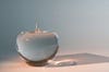
Quantum Apple

Robots
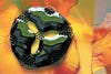
Eleanor Rigby
Lichen Colony
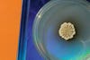
Bacillus subtilis

Solar Cell

Pin toy
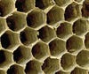
Hornets’ Nest

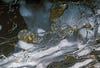
Water
Ferrofluid