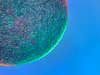
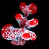
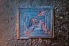
18th place. Table salt crystal. Magnified 10x. Saulius Gugis/Nikon Small WorldSHARE
Mouse brains and butterfly wings. Tick heads and snowflakes. It’s that time of the year again, when winners of the Nikon Small World photomicrography contest are announced. Running 47 years strong and open to all, it’s considered the premiere set of awards for folks with an interest in both microscopy and photography—the Oscars of the nearly-invisible world if you will.
Small World entries
This year, Nikon received almost 1,900 entries to the Small World contest, from 88 different countries. The majority of the winners come from the academic world, but not all: Some are simply amateur photomicrographers with a passion for the teeny tiny.
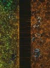
Insects were by far the most popular subject among the winning crowd. But it’s the microscopic images of everyday stuff, like Saulius Gugis’ 10x magnified table salt crystal (above), that intrigue us the most. Felice Placenti’s image of cotton fabric with pollen grains, also magnified 10x, was another staff favorite (check it out below).
Techniques
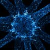
Contestants employed a variety of techniques to tease out the details in their photos. The use of image stacking and fluorescent light, as well as various dyes, proved popular. The overall winning image, shown below, is actually a combination of 200 different shots—magnified 60x—and captured through a custom-built microscope.
Our favorite images
Which is your favorite image from the Nikon Small World contest? Scroll through and let us know in the comments below! And be sure to check out the winners of Nikon’s Small World in Motion video contest as well. You can also view past Small World contest winners here.
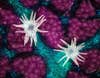
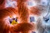
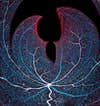
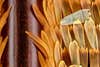
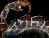
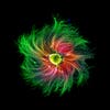
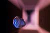
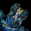
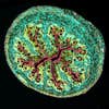
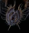
1st place. Trichome (white appendages) and stomata (purple pores) on a southern live oak leaf. Magnified 60x. Jason Kirk, Baylor School of Medicine/Nikon Small World13th place. Cotton fabric with pollen grains. Magnified 10x. Dr. Felice Placenti, FP Nature and Landscape Photography/Nikon Small World11th place. Vasculature of a mouse retina. Magnified 20x. Jason Kirk & Carlos P. Flores Suarez, Baylor College of Medicine/Nikon Small World10th place. Vein and scales on a butterfly wing (Morpho didius). Magnified 20x. Sébastien Malo/Nikon Small World 3rd place. The rear leg, claw, and respiratory trachea of a louse (Haematopinus suis). Magnified 5x. Frank Reiser, Nassau Community College/Nikon Small World 17th place. Filamentous strands of Nostoc cyanobacteria captured inside a gelatinous matrix. Magnified 4x. Martin Kaae Kristiansen, My Microscopic World/Nikon Small World4th place. Sensory neuron from an embryonic rat. Magnified 10x. Paula Diaz, MinusPain, Pontificia Universidad Catolica de Chile/Nikon Small World 19th place. Calcite crystal inclusion suspended in a spinel gemstone. Magnified 40x. Billy Hughes, Lotus Gemology/Nikon Small World12th palce. Breast organoid showing contractile myoepithelial cells (blue) crawling on secretory breast cells (red). Magnified 40x. Jakub Sumbal, Masaryk University/Nikon Small World 7th place. The head of a tick. Magnified 10x. Tong Zhang & Paul Stoodley, Ohio State University/Nikon Small World8th place. Cross section of mouse intestine. Magnified 10x. Dr. Amy Engevik, Medical University of South Carolina/Nikon Small World5th place. The proboscis of a housefly (Musca domestica). Magnified 40x. Oliver Dum, Medienbunker Produktion/Nikon Small World16th place. An in vivo snapshot of the neurons surrounding the mouth and tentacles of a juvenile starlet sea anemone (Nematostella vectensis). Magnified 20x. Ruohan Zhong Stowers, Institute for Medical Research/Nikon Small World2nd place. A microfluidic device containing 300,000 networking neurons in 2 isolated populations. Both sides were treated with a unique virus and bridged by axons. Magnified 40x. Esmeralda Paric & Holly Stefen, Dementia Research Centre, Macquarie University/Nikon Small World18th place. Table salt crystal. Magnified 10x. Saulius Gugis/Nikon Small World