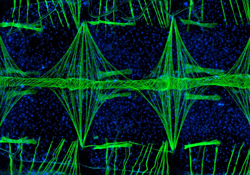
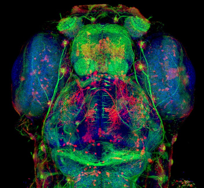
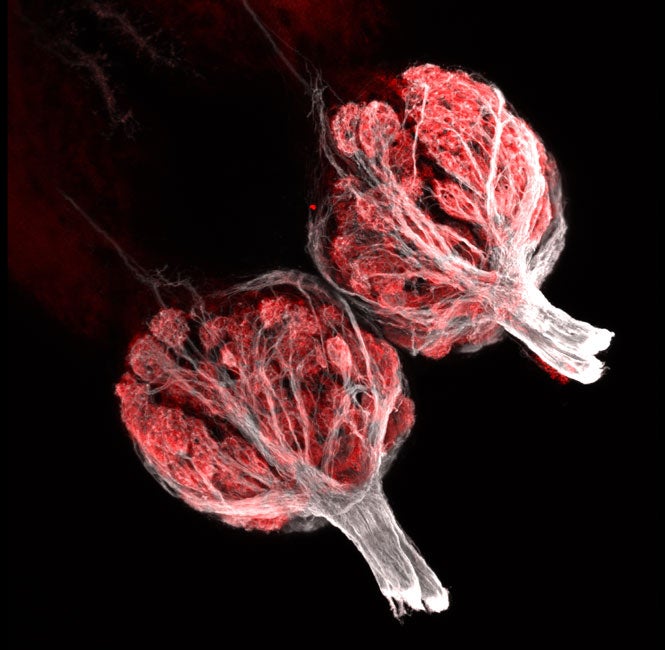
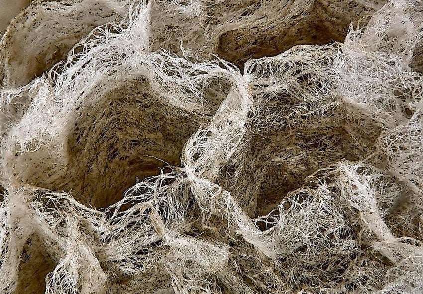
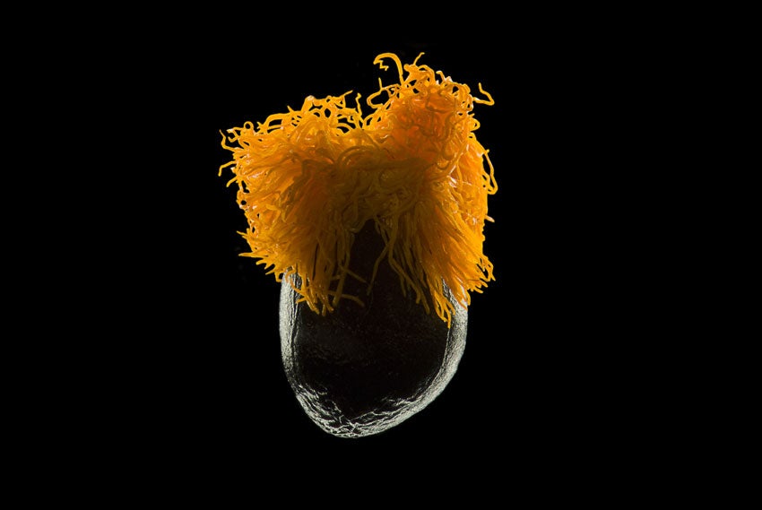
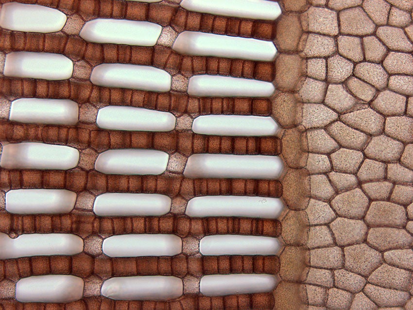
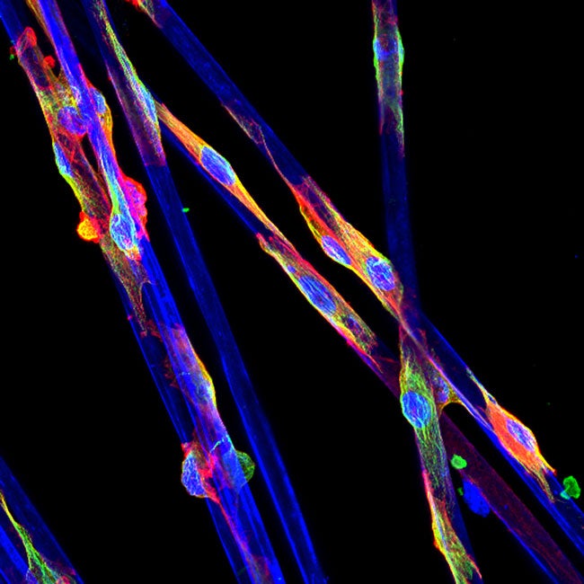
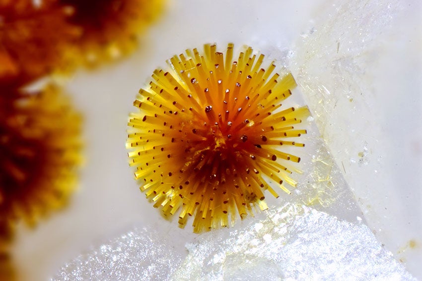
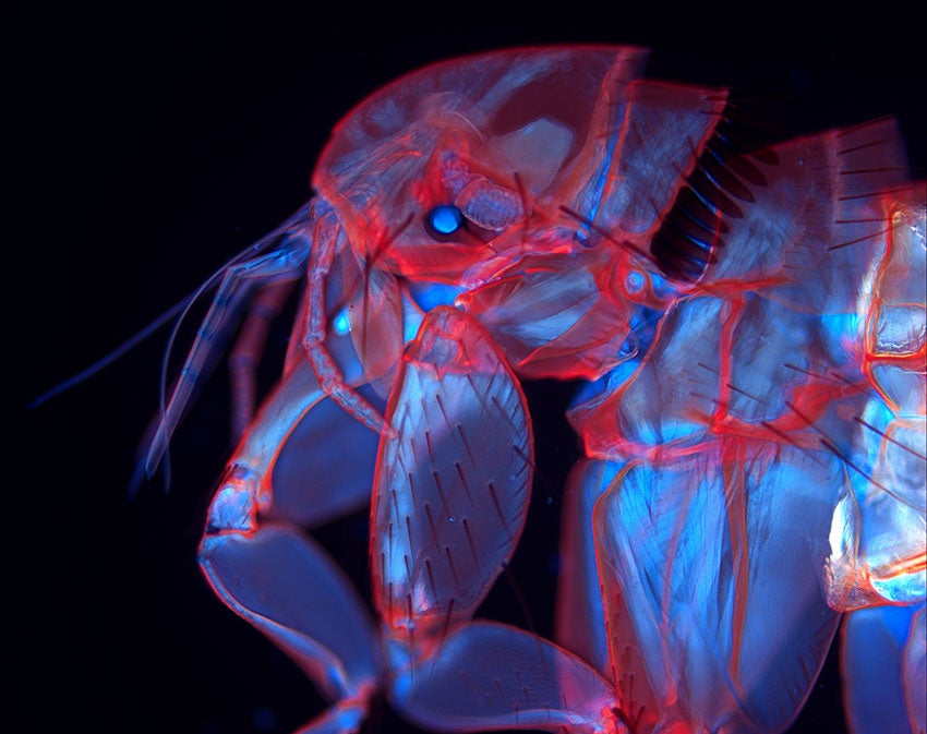
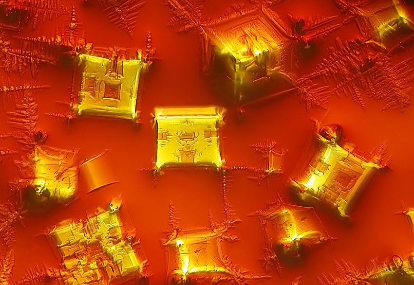
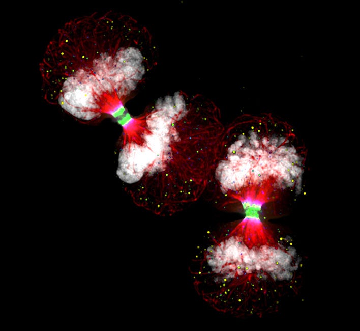
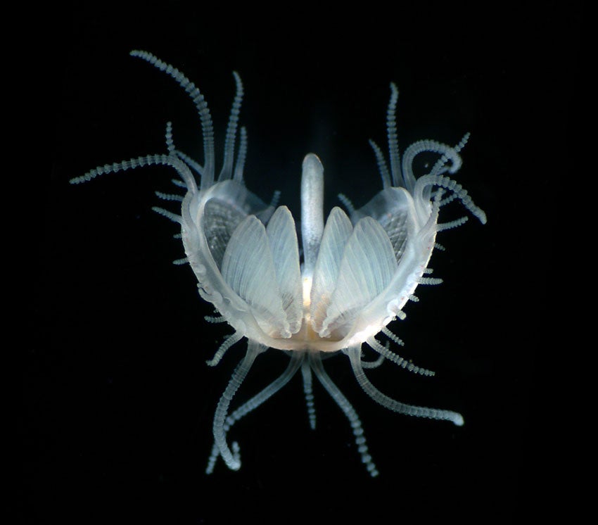
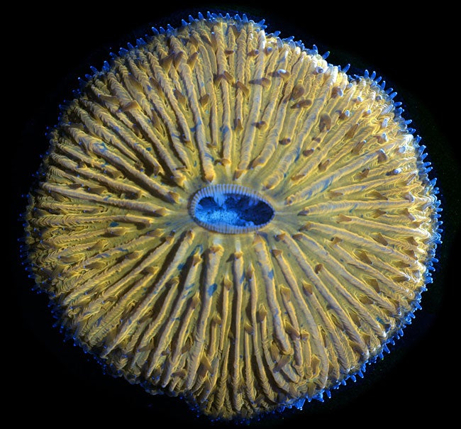
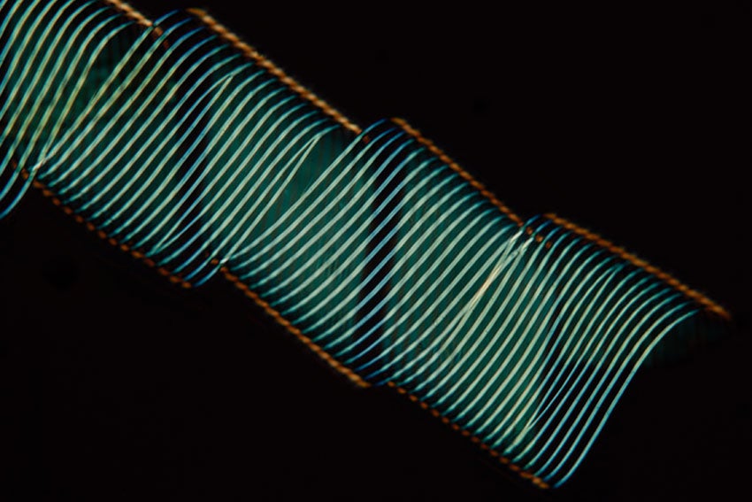
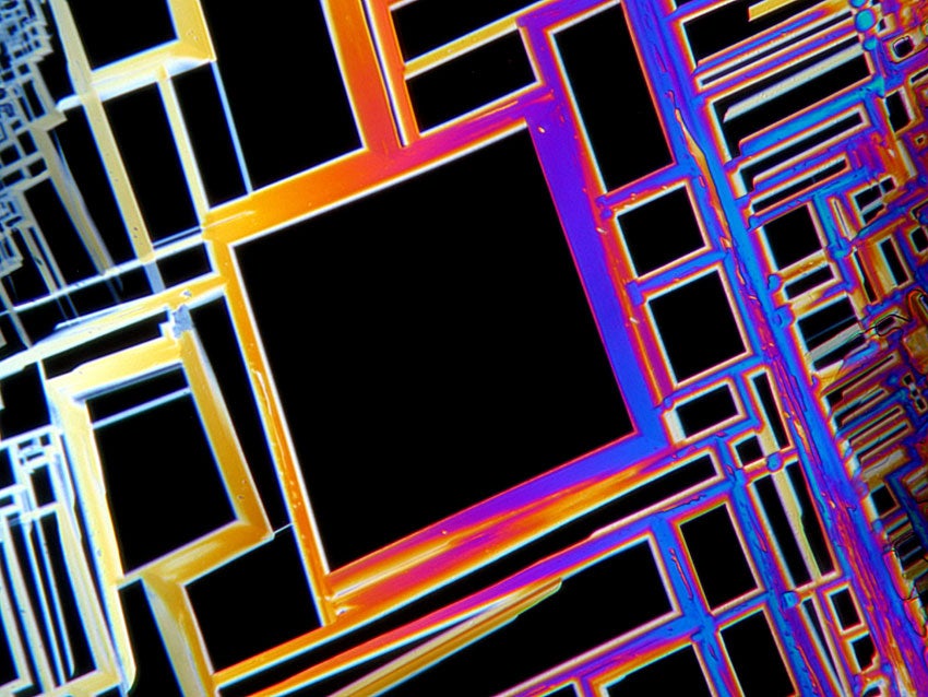
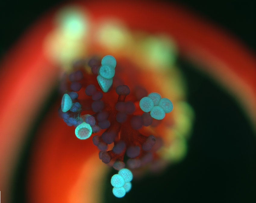
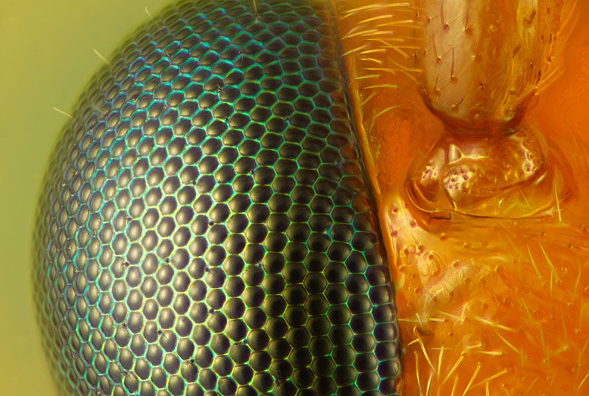
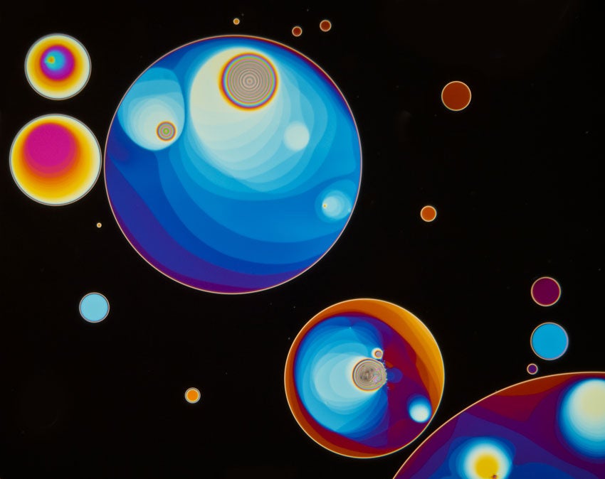
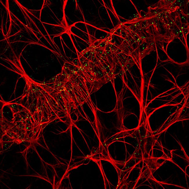
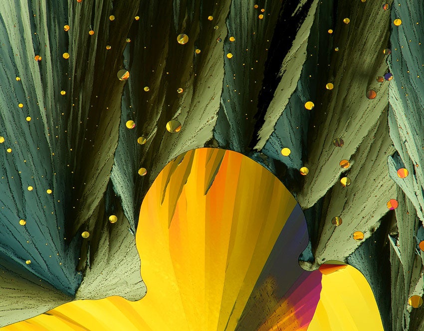
Nikon has announced the winners of their 36th annual Small World** c****ontest**, which collects best images captured through a microscope over the course of the year. The results are often as surreal as they are beautiful. Subjects range from tiny animal organs completely invisible to the naked human eye. A board of judges, which included journalists and scientists, whittled down more than 2,200 entries into this incredible 20 image gallery. **1st Place: **Jonas King Anopheles gambiae (mosquito) heart (100X) Fluorescence Vanderbilt University, Department of Biological Sciences, Nashville, Tennessee, USA**2nd Place: **Dr. Hideo Otsuna 5-day old zebrafish head (20X) Confocal University of Utah Medical Center, Department of Neurobiology and Anatomy, Salt Lake City, Utah, USA**3rd Place: **Oliver Braubach Zebrafish olfactory bulbs (250X) Confocal Department of Physiology & Biophysics, Dalhousie University, Halifax, Nova Scotia, Canada4th Place: Riccardo Taiariol Wasp nest (10X) Extended Depth of Field Stereomicroscopy La Spezia, SP, Italy**5th Place: **Viktor Sykora Strelitzia reginae (bird of paradise) seed (10X) Darkfield Institute of Pathophysiology, First Medical Faculty, Charles University, Prague, Czech Republic6th Place: Dr. John Huisman Martensia sp. (red seaweed), living specimen (40X) Brightfield Murdoch University, School of Biological Sciences and Biotechnology, Murdoch, Western Australia, Australia7th Place: Yongli Shan Endothelial cell attached to synthetic microfibers, stained with microtubules, F-actin and nuclei (2500X) Fluorescence, Confocal The University of Texas Southwestern Medical Center, Dallas, Texas, USA**8th Place: **Honorio Cocera-La Parra Cacoxenite (mineral) (18X) Reflected light Geology Museum, University of Valencia, Benetusser, Valencia, Spain**9th Place: **Dr. Duane Harland Ctenocephalides canis (flea) (20X) Fluorescence AgResearch Ltd. Lincoln, New Zealand**10th Place: **Yanping Wang Crystallized soy sauce (16X) Reflected and Transmitted Light Beijing Planetarium, Beijing, China**11th Place: **Dr. Paul D. Andrews Telophase HeLa (cancer) cells expressing Aurora B-EGFP (green) (100X) Deconvolution University of Dundee, Dundee, Scotland, UK**12th Place: **Dr. Gregory Rouse Juvenile bivalve mollusc, Lima sp. (10X) Darkfield Scripps Institution of Oceanography, La Jolla, California, USA**13th Place: **James Nicholson Orange Fungia (mushroom coral), live specimen (166X) Fluorescence NOAA NOS NCCOS Coral Culture and Collaborative Research Facility, Charleston, South Carolina, USA**14th Place: **Dr. Stephen Lowry Spiral vessels from banana plant stem (32X) Polarized light University of Ulster, Portstewart, Co. Londonderry, UK**15th Place: **Dr. Ralf Wagner Divaricatic acid from Evernia divaricata (lichen), recrystallized from acetone (10X) Polarized light Düsseldorf, Germany**16th Place: **Dr. Robert Markus Mirabilis jalapa (four o’clock flower) stigma with pollen (100X) Epifluorescence and 3D reconstruction Institute of Genetics, Biological Research Center of the Hungarian Academy of Sciences, Szeged, Hungary**17th Place: **Charles Krebs Ichneumon wasp compound eye and antenna base (40X) Reflected (Episcopic) Light Illumination Charles Krebs Photography, Issaquah, Washington, USA**18th Place: **Gerd Guenther Soap film (150X) Incident Brightfield Düsseldorf, NRW, Germany**19th Place: **Cameron Johnson Wistar rat retina outlining the retinal vessel network and associated communication channels (100X) Confocal The University of Auckland, Auckland, New Zealand**20th Place: **Dr. John Hart Crystallized melt of sulfur and acetanilide (10X) Transmitted Light, Crossed Polars Hart3D Films and Dept. Atmospheric and Oceanic Sci. Univ. Colorado, Boulder, Boulder, Colorado, USA